ONSITE NUCLEAR VIDEO IMAGING
Christophe LE GOALLER, Jean-Raymond COSTES
Commissariat à l'Energie Atomique (CEA)
Rhône Valley Research Center
BP 171, 30207 Bagnols-sur-Cèze Cedex (France)
ABSTRACT
This reports presents the most recent development worked out by the Nuclear Facility Decommissioning Unit of CEA, in the field of in-situ nuclear imaging.
The latest device of video gamma camera "Aladin 2" is briefly described, and the perfomance reached by this prototype are summarized. Some images obtained in decommissioning cells during in-situ qualification are displayed.
The second part of the report focuses on the first prototype of video alpha camera. The basic principle is explained, and the latest results of laboratory or in-situ trials are commented.
INTRODUCTION
Satisfactory completion of decommissioning operations requires precise knowledge of the radiological condition of the facilities. Without reliable basic data, it is difficult to adapt the operating conditions to the site requirements.
Radiological balances are currently based on discrete measurements obtained from probes that provide crude maps based on irradiation values. Gamma spectrometry devices can also be used to identify the major g -emitting radionuclides. However, except in very simple environments, it is difficult to obtain accurate information concerning the location or extent of hot spots. Although widely used and in constant progress in many fields (nondestructive examination, medical diagnostics, scientific research, etc.), imaging techniques are rarely applied in nuclear facilities.
For nearly a decade, the nuclear facilities decommissioning unit of the Commissariat à l'Energie Atomique has been developing and implementing radiation imaging systems suitable for site work, combining radioactivity display images with quantitative measurement data.
GAMMA IMAGING
Historical Background
Gamma imaging is not new, and is widely used for medical purposes. Nevertheless, the first prototype unit capable of displaying the distribution of hot spots by superimposing a "gamma image" on a photographic emulsion, based on the pinhole camera concept, was developed only in the late 1980s [1]. The first video gamma camera designed for rapid localization of gamma activity in nuclear facilities, the Aladin prototype, appeared in 1993[2]. This patented device addressed the requirements of in situ work at nuclear decommissioning sites: compact design, interactivity (a few minutes are sufficient to obtain an image) and acceptable sensitivity (sources generating a dose rate of 1 m Gy· h-1 can be localized in 5 minutes). Recently a new prototype, Aladin 2, with improved performance and better suited for industrial applications, has been developed and is now being implemented in the field.
Description
The gamma camera uses a double-cone collimator to form a visible-light or gamma image in the same way as a pinhole camera. Unlike the initial design, the image is not formed on a photographic emulsion, but on a scintillator: a CsI(Tl) disk 4 cm in diameter and generally between 2 and 4 mm thick. Gamma photons interact in the scintillator to generate luminous photons with a wavelength of 550 nm, forming an image. As in medical imaging systems, the scintillator output signal is amplified and collected on a CCD array. We use a second-generation image intensifier tube with a double microchannel plate, and a large diameter (40 mm) entrance window. The CCD array measures less than a centimeter on a side, and is therefore coupled to the tube screen via a tapered fiber-optic demagnifier. The unit is compact (12 cm in diameter and 30 cm long) and is protected against spurious radiation by Denal tungsten alloy shielding 2 cm thick, ensuring 85% attenuation at 1.25 MeV and 95% at 660 KeV. A color video camera is secured to the shielding.
The gamma camera and color camera signals are transmitted to video acquisition cards in a PC-compatible microcomputer running the imaging software.
Utilization
The Aladin 2 prototype can be operated in two modes: hot-spot detection or localization.
The sensitivity of the Aladin 2 device allows "real-time" detection of hot spots (i.e. within 5 seconds). In this mode, the image acquisition is performed entirely on the CCD array, which accumulates the incident signals for a predetermined time (t = k ´ 40 ms); this is equivalent to setting the exposure time of a conventional camera. The resulting accumulated signal is transmitted to the PC video acquisition card; the screen displays an 8-bit image updated every k ´ 40 ms.
The facilities examined are often large and complex, and the hot spot locations are not known beforehand. The "detection" mode allows the camera to sweep the field of view while the PC monitor displays the real-time color video image and the periodically updated gamma image. A hot spot results in signal saturation, producing a "white spot" that is clearly distinguishable from the noise; it does not provide precise information on the source location, but is conclusive evidence of the existence of an irradiating zone.
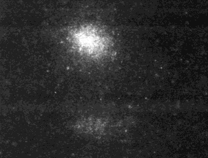
The least favorable case would be a facility in which the hot spot dose rate at the camera distance is very low, on the order of 10 m Gy.h-1 (1 mrad· h-1). Under these conditions the CCD integration time is 5 seconds, requiring slow video sweeping. In most case, however, the irradiation levels generated by hot spots range from 1 mGy.h-1 (100 mrad· h-1) to 1 Gy.h-1 (100 rad· h-1) at the camera position: detection is possible in only a few tenths of a second or even in real time (40 ms). Figure 1 shows the image corresponding to in real time of a hot spot irradiating a around 0,4 Gy.h-1 at the camera position.
Figure 1. Real time detection of a hot spot - Dose rate received by the camera : 0.4 Gy.h-1.
Once a hot spot has been detected, an 8-bit dynamic range is rarely sufficient to localize the source without accumulating a large number of incident images. The video acquisition card is used to integrate successive CCD signals (generally in real time) into a 16-bit image; white spots appear on the display with increasing intensity during the integration period. The acquisition time ranges from 5 seconds to 10 minutes, depending on the irradiation level and on the intensifier tube gain. Figure 2 shows a typical integrated gamma image of a highly irradiating source with a camera-position dose rate of 4 Gy.h-1.
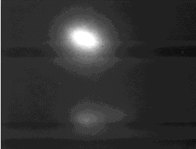
Figure 2. 16-bit gamma image of a highly irradiating source - Acquisition time : 5 s.
After low-pass filtering and color enhancement, the gamma image is superimposed on the corresponding visible-light image to localize the hot spots in the field of view. Parallax errors are corrected by the software during processing by a calibration sequence (the gamma camera shutter is opened to obtain a black-&-white visible-light image that is compared with the color image from the external video camera to ensure accurate superimposition). The result is shown in Figure 3.
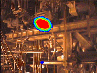
Figure 3. Superimposed video and 16-bit gamma images
Performance
In its present configuration, the device is capable of detecting point sources irradiating a 10 m Gy.h-1 (1 mrad.h-1) dose rate, in 5 seconds (8-bit image depth). Localization of a source generating 0.5 m Gy· h-1 (0.05 mrad.h-1) requires a 10-minutes exposure (Signal to Noise ratio » 2). The photon flux is very weak for low-level irradiation sources : for example, a dose rate of 10 m Gy· h-1 (1 mrad· h-1) generated by 60Co photons (1.25 MeV), corresponds to a photon flux of 520 ph· cm-2s-1. The limiting factors are the aperture diameter of the collimator (1.4 mm is used to obtain an angular resolution of a few degrees), and the poor interaction probability (4.4 ´ 10-2 interaction per photon for 1.25 MeV photons trough a 2 mm CsI(Tl) scintillation crystal). The detection sensitivity can be improved ,at the expense of the resolution, through the use of collimators with a larger aperture. Another option would be to use a thicker scintillator crystal to increase the interaction probability.
The angular resolution varies according to the photon energy flux, the aperture diameter, the scintillator thickness and the collimator solid angle. The present configuration (1.4 mm aperture, 2B4 mm thick scintillator, 50E collimator angle) is capable of discriminating 60Co sources with an angular separation of less than 5E (the separability criterion assumes 30% relative contrast between the two peaks). In the most favorable case (cesium) the resolution is about 2.5E. An effective way of enhancing the resolution is to diminish the collimation angle: a 30E collimator provides an angular resolution of 1.3E at the same energy, but with a corresponding reduction in the field of view.
Detection of hot spots in a few seconds can be useful not only in decommissioning a nuclear facility, but also during routine operation or maintenance, for instance to follow a moving source. Localizing irradiating source in a few minutes enables multiple views from different angles to be observed, for accurate localization, especially in complex environments.
After precise calibration, it is possible to estimate the dose rate generated by a punctual source at camera position. As a matter of fact, the pixel size reached after a given integration time depends on the dose rate received (for given amplification gain and photon energy). The knowledge of dose rate 1 meter apart from the source requires the use of a telemeter. Once the source has been localized, more precise data on the source (accurate dose rate, gamma spectrum) may also be obtained by the use of a strong collimated separate probe.
ALPHA IMAGING
Work has recently begun on the development of an alpha video camera intended for remote localization of a -emitting hot spots, with potential applications in fuel reprocessing or radiochemistry facilities.
Principle
The patented[3] design is based on detecting scintillation of the air excited by alpha particles in the 220-520 nm spectrum band, and particularly (60%) between 320 and 390 nm. This phenomenon is due to excitation of the nitrogen molecule: 50% molecular excitation, 45% ionization or charge transfer with excitation, and 5% atomic excitation. Argon also exhibits excellent emission properties, but is present in such small quantities that its contribution to the luminescence of the air is negligible. Oxygen is not a fluorescence emitter, but instead has a quenching effect on the excitation. Air thus has a very poor scintillation yield, some five times lower than pure nitrogen. An a particle with an energy of 5.48 MeV emits about 1000 photons in nitrogen, and fewer than 200 in air.
This photon flux can be focused by a Fresnel lens and collected on a CCD array with a spectral response extending into the near ultraviolet. A slow scan cryogenic CCD is used, with a high quantum efficiency and large pixels (24 x 24 m m). It supplies a 16-bit image to a video acquisition card in a PC.
Results
Strongly contaminated a sources can be localized in a relatively short time: only one minute was necessary to obtain an "alpha" image of an americium-241 source with a contamination level of 100 MBq (3.3 MBq.cm-2) at a distance of over four meters (S/N ratio » 4). This result was obtained in darkness with a relatively narrow field of view (6° solid half-angle). At the detection limit, a plutonium point source generating 1350° Bq over 1° cm2 at a distance of 4° m can be hardly detected, through a 5 mm plexiglas wall, in 30 minutes. The same source is easily detected at a shorter distance, 1.2 m, without plexiglas screen, in 15 minutes (S/N ratio » 9).
Tests also showed that alpha sources can be observed through Plexiglas, triplex or polycarbonate screens of limited thickness. The attenuation coefficients of these three materials were found to range from 0.85 to 0.97 per millimeter.
The results of a test under realistic conditions (a telemanipulator tong observed from a distance of 1.25 m through a 5 mm Plexiglas glove box) are shown in Figure 4. The major contaminants were Curium isotopes at levels of around 1 MBq· cm-2, allowing short acquisition times on the order of five minutes. The most severely contaminated areas are clearly visible: mainly the inside of the jaws and a few edges or asperities around the body.
It is interesting to observe the area between the jaws, where scattered and attenuated points are visible. This may be attributable to the medium-level energy (about 6 MeV) of the a particle emitted by the curium isotopes. Alpha particles follow straight-line paths in air, with a mean stopping power of 1.3 MeV$cm-1. Thus the range of alpha radiation with an energy of 6 MeV reaches 4.3 cm, compared with the 8 cm gap between the jaws.
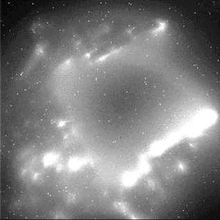
Figure 4. Image of a
Curium-contaminated telemanipulator tong
(5-minutes acquisition time - Camera to subject distance: 1.25 m - Plexiglas
screen thickness : 5 mm)
CONCLUSION
Alpha imaging is a promising development; it is too early to speak of an industrial prototype at this time, but the feasibility of the concept has been demonstrated, and first in-situ qualification results are promising.
Although gamma imaging is not yet widely used for in situ operation in nuclear facilities, our prototypes are increasingly used at decommissioning sites and are envisaged for other sectors such as maintenance work in nuclear power stations.
Many in situ gamma imaging devices are now being developed. Most of them are based on "indirect" conversion of gamma photons to visible-light photons generating an electrical signal. Two options are available for gamma photon collimation : a coded mask provides good sensitivity, while a double-cone "pinhole" collimator results in a compact design and can be adjusted according to specification requirements (sensitivity, resolution, field of view).
It is highly likely and desirable that these multiple developments will make in situ imaging a common application in nuclear facilities.
REFERENCES
1. G. SIMONET, "Télétopographie Gamma", 6xt Symposium ASTM/EURATOM on Reactor Dosimetry, Jackson Hole, USA, june 1987.
2. G. IMBARD, H. CARCREFF "Development of a prototype gamma camera for use in decommissioning facilities", Waste Management 1995, Tucson, Arizona, march 1995.
3. G. IMBARD, JF PINEAU, brevet français 96.03976.